RAMAN SPECTROMETER
DOWNLOAD REQUEST FORM HERE
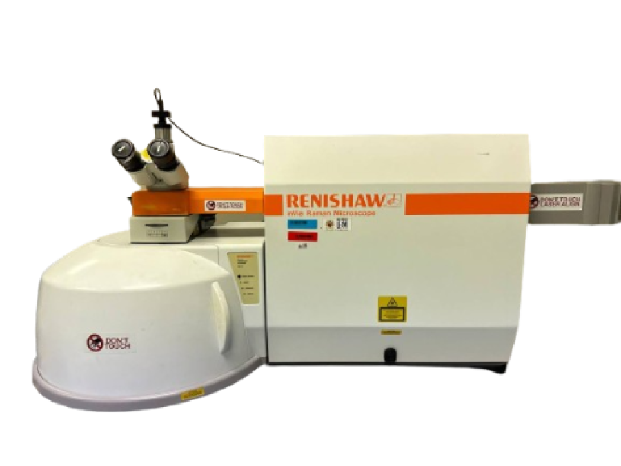
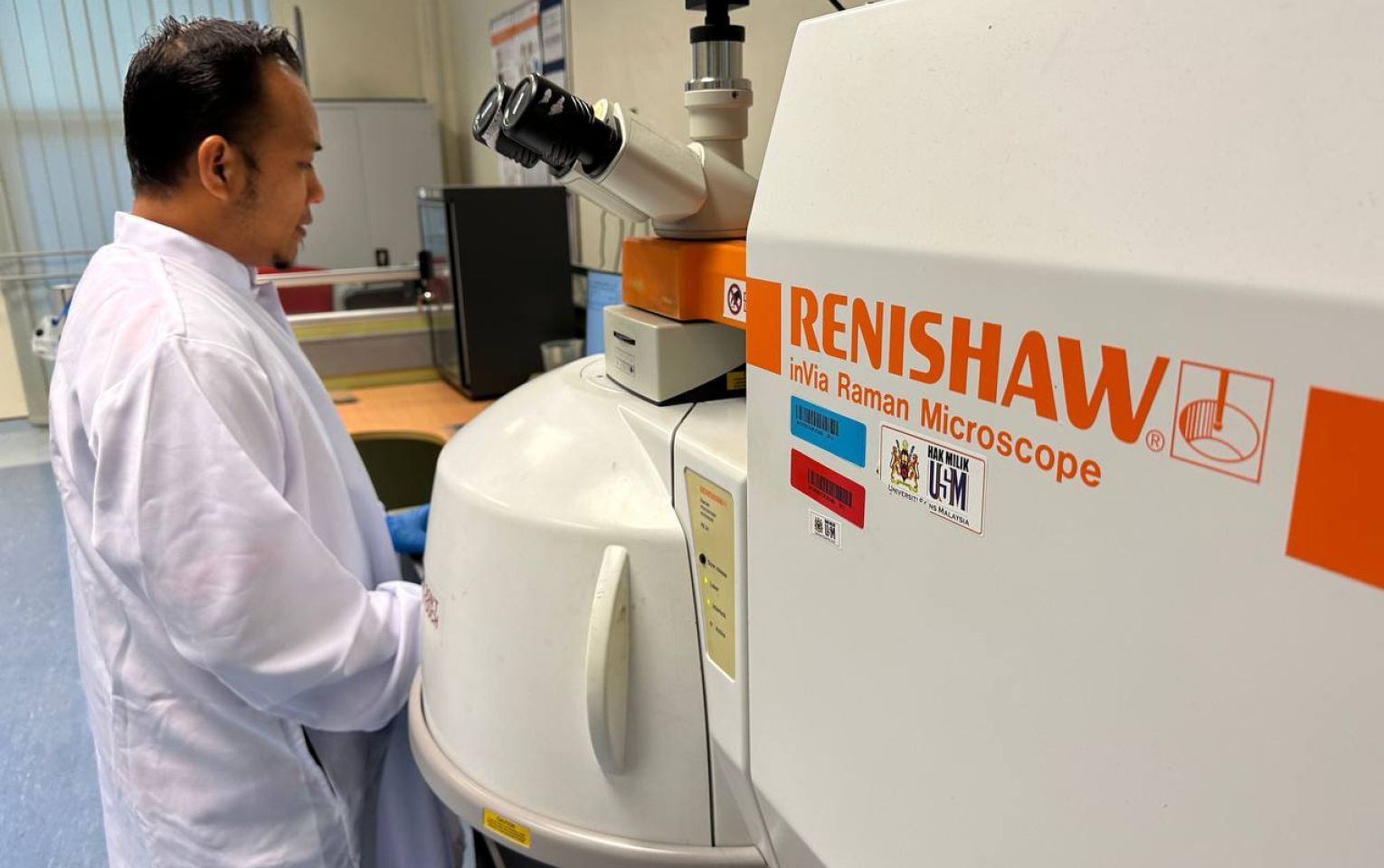
INSTRUMENT NAME & MODEL: Raman Spectrometer (Renishaw inVia Raman Microscope)
AVAILABLE LASER: 455nm, 532nm, 633nm and 785nm
Micro-Raman imaging spectroscopy is a useful technique to identify structural fingerprint and compound of films, powder, solids or liquids samples by measuring the molecular vibrations. This instrument enables to measure several types of analyses such as point measurement, depth profiling (ranging thickness from 2 to 15 μm) and also can do mapping.
This technique is a straightforward, non-destructive and requiring no sample preparation. Raman spectroscopy involves illuminating a sample with monochromatic light and using a spectrometer to examine light scattered by the sample. Principle of this technique is based on inelastic scattering of monochromatic light, usually from a laser source. Inelastic scattering means that the frequency of photons in monochromatic light changes upon interaction with a sample. Photons of the laser light are absorbed by the sample and then reemitted. Frequency of the reemitted photons is shifted up or down in comparison with original monochromatic frequency, which is called the Raman effect. This shift provides information about vibrational, rotational and other low frequency transitions in molecules.
CATEGORY: SPECTROSCOPY LABORATORY

Special Features
- Excitation : 633 nm HeNe laser (20mW)
- Grating : 1800 g/mm (1cm-1)
- Laser Power : 10% (required to assure sample integrity)
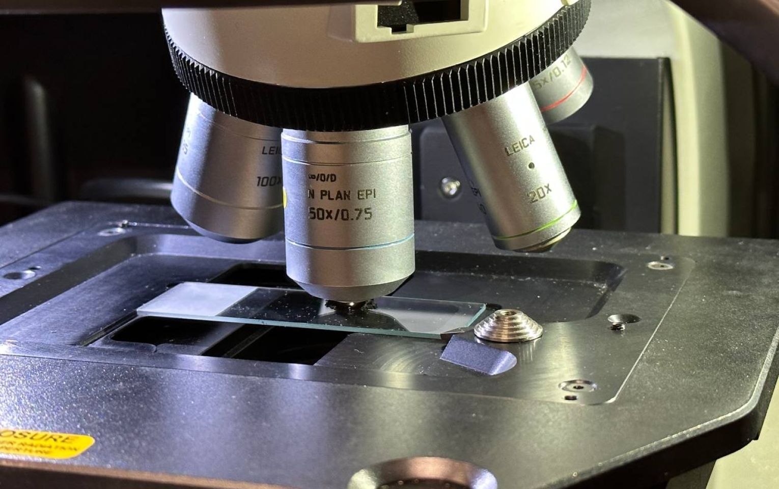
- Objective : 50x N plan (1um laser spot size)
- Spectral Range : SynchroScanTM 100 cm-1 to 4000 cm-1

Applications
- Evaluate the quality of materials such as Graphene/CNT materials.
- Measure depth profiling of multi-layered component such as polymers samples.
- Identify and discriminate between different types of samples from large amounts of sample to residues.
- Characterized the distribution of active ingredients such as in pharmaceutical tablet.
- Identified inclusions in geological samples.
- Identify the spatial distribution and characteristic spectra of biomolecules materials.
- Quality of the molecular bond structure.
- Vibration mode of metal oxide.
- Investigate the chemical composition of historical documents
- Single walled carbon nanotubes (SWCNTs)
- Purity of carbon nanotubes (CNTs)
- Electrical properties of carbon nanotubes (CNTs)
- sp2 and sp3 structure in carbon materials
- Diamond like carbon (DLC) coating properties
- Defect/disorder analysis in carbon materials
- Diamond quality and provenance
- Characterization of intrinsic stress/strain
- Purity
- Alloy composition
- Superlattice structure
- Defect analysis
- Powder content and purity
- Raw material verification
- Crystallinity
- In vivo analysis and skin depth profiling

Sample Requirements
- Powder
- Film
- Solid
Typical Results
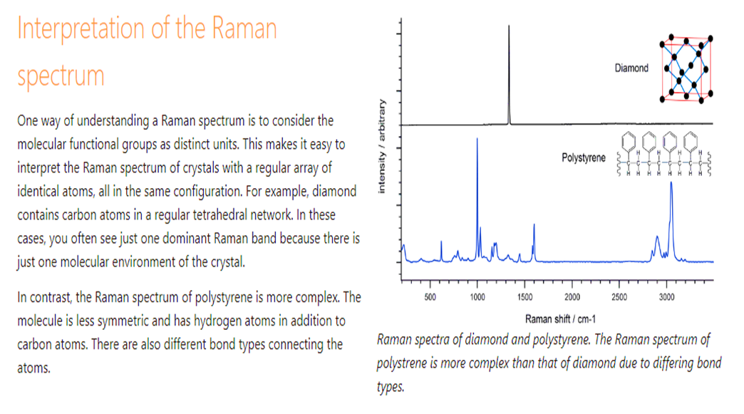
RAMAN Typical results


