XHR-FESEM-Extreme High Resolution Field Emission Scanning Electron Microscope
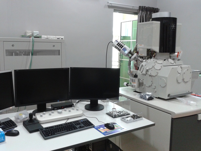
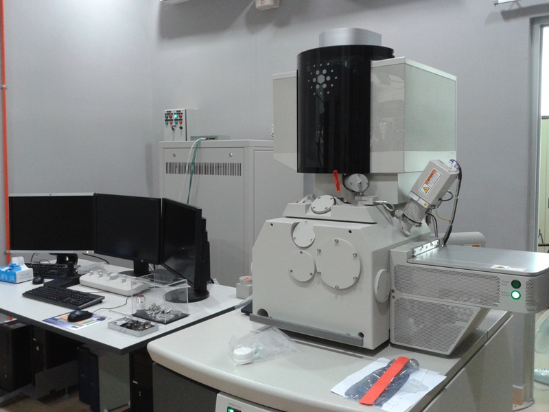
Instrument Name & Model: XHR Extreme High Resolution Field Emission Scanning Electron Microscope (XHR-FESEM) Model FEI Verios 460L
Special Features:
Electron beam resolution:
Resolution @ optimum WD
• 0.6 nm at 30 kV (STEM *)
• 0.7 nm at 15 kV
• 0.7 nm at 1 kV
• nm at 500 V (ICD **)
• 1.2 nm at 200 V (ICD **)
Landing energy range
• 20 eV - 30 keV
Probe current
• E-beam: 0.8 pA up to 100 nA
Detectors
• In-lens SE detector (TLD-SE)
• In-lens BSE detector (TLD-BSE)
• In-column SE detector (ICD) **
• In-column BSE detector (MD) **
• Everhart-Thornley SE detector (ETD)
• IR camera for viewing sample/column
• Chamber mounted Navigation Camera
• Retractable low voltage, high contrast solid-state backscatter electron detector (DBS)
• Oxford Silicon Drift Detector (SDD) - X-Max EDS/EDX detector
Sample sizes
• Maximum size: 0.5cm
• Maximum size: 1cm (Mold sample)
• Maximum sample thickness (via loadlock/chamber door): 19 mm incl. stub
• Weight: 200 g (incl. holder)
Sample holders
• Multi-stub holder (5 stubs)
• Multi-sample cross-sectional holder
• Single stub mount, mounts directly onto stage
• Various wafer and custom holder(s) available by request
Applications:
This instrumentation is particularly suitable for the study and characterization of:
• Biological materials (Dry Sample Only)
• Polymeric materials
• Metallic materials
• Composite materials
To get information on the morphological, chemical and analytical study of materials.
*Samples required : Dry & Non Magnetic
Location: Microscope Room (G045)
Contact:
ELECTRON MICROSCOPY LAB
Universiti Sains Malaysia
14300 Nibong Tebal, Penang, Malaysia